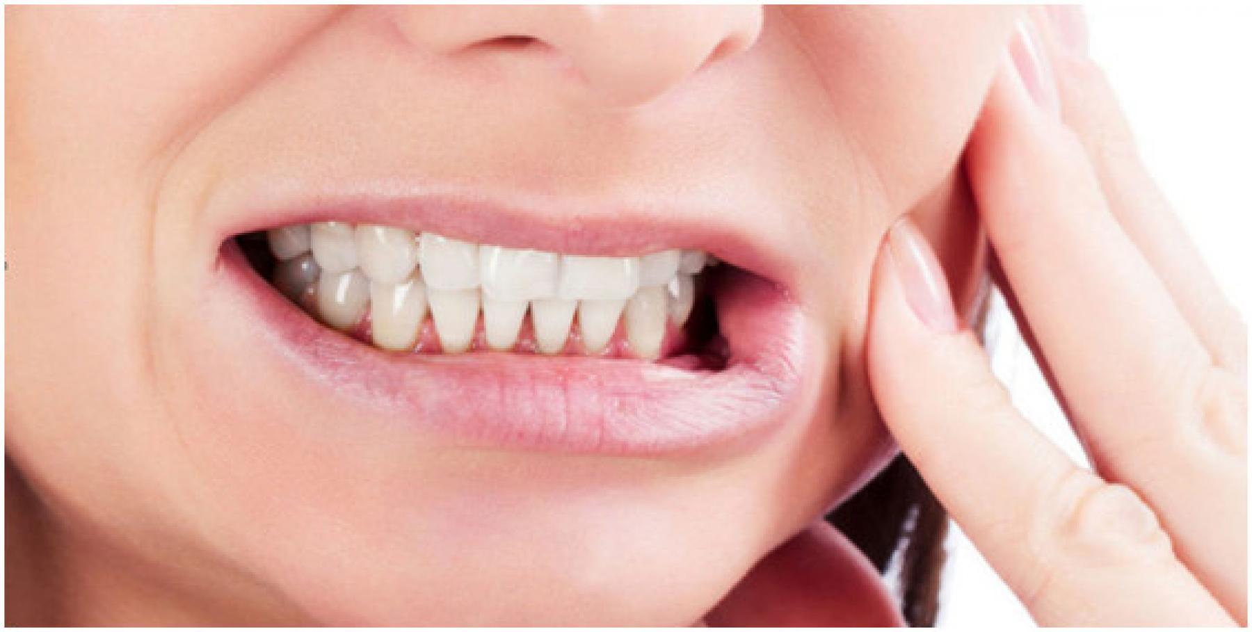
Bruxism or “teeth grinding” is a complicated phenomena which has a prevalence or 8%-31.4% in the general population (Manfredini et al. 2013). In the last 5 years, the classification and definition of bruxism has changed. An International Consensus of Dentistry Experts (International Network for Orofacial Pain an related Disorders Methodology; INfORM), the German Deutsche Gesellschaft für Funktionsdiagnostik und -Therapie in der Zahn-, Mund- und Kieferheilkunde (DGFDT), and the Deutsche Gesellschaft für Zahn-, Mund- und Kieferheilkunde (DGZMK)) (2019) have collaborated to create a strong international consensus about the etiology, assessments and management of bruxism.
Bruxism is a disease which derives predominantly from the masticatory muscles
Bruxism is not a disease but a collective term for abnormal mouth habitual activity together with increased jaw-muscle activity (Lobbezoo et al. 2018). The complexity of the neurophysiological mechanism has been poorly understood until now. Risk factors (anatomical, psychosocial, and/or neuromuscular) are complex, and the interference of the different risk factors is controversial (Manfredini et al. 2013).
Bruxism belongs to the sub-classification of myofascial TMD (Axis I, DC/TMD consortium)
The general belief of physical therapists is that bruxism is a kind of myogenic TMD which is increased by stress, however, this is not the case. TMD, such as difficulty closing and opening the mouth, jaw locking, temporomandibular joint sounds, and a feeling of stiffness or jaw fatigue can be a possible risk factor for bruxism (Ciancaglini et al. 2001). Feelings of stiffness or jaw fatigue are more frequently seen in sleep bruxism; whereas, awake bruxism is more associated with joint sounds and depressive mood (Selms et al. 2004; Manfredini et al. 2013). A recent study confirmed that the prevalence of bruxism is increased when there is an increased severity of TMD with increased neck disability and pain (von Piekartz 2019).
Bruxism is predominantly peripherally activated and therefore, it has a dominant input trigger
Palpation of masticatory muscles, especially the masseter and temporal muscles, may be significantly sensitive in bruxers but is it fair to hypothesize that there is a causal relationship between hypersensitivity, increased muscle tone, and the bruxers’ complaints? In patients with bruxism, these signs are mainly centrally mediated, not peripherally. There is convincing evidence, however, that (sleep-related) bruxism is part of an arousal response. Disturbances in the central dopaminergic system have been implicated in the etiology of bruxism as well. Furthermore, there is a role for factors like smoking, alcohol use, diseases, trauma, and heredity, while the proposed role of stress and other psychological factors is probably smaller than previously assumed (Lobbezoo and Naeije et al. 2001, Lobbezoo et al. 2018 ).
Bruxism and Bracing are two different phenomena
Most physical therapists are convinced that bruxism is grinding during the night and bracing is different from clenching during the day. This is the wrong premise. Somnography and polysomnography studies have shown that clenching and grinding are (unconscious) mechanisms with teeth contact during the day or at night (Raphael et al. 2013, Muzalev et al. 2017).
Posture changes, especial forward head posture, cause an increased masticatory system activity and leads to bruxism
From different studies, it is known that there is no clear evidence for the existence of a predictable relationship between occlusal and postural features. It is clear that the presence of bruxism (and also of TMD) is not related to the existence of measurable occlusal-postural abnormalities in adults (Cesar et al. 2006, Manfredini et al. 2012 ). In children, head posture, TMD, craniofacial morphology, and bruxism seems to be minor to moderately associated with each other (Kritsineli et al. 1992, Motta et al. 2011).
Bruxers need a splint from the dentist because the bite is a causal factor
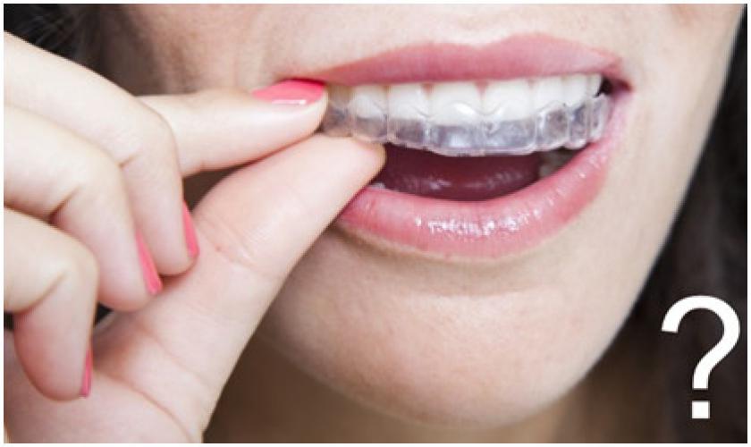 Patients and therapists are often influenced by the media or by non-scientific, commercial publications that a splint may help to get rid of (the complaints caused by) bruxism. Based on several reviews, there is no evidence what-so-ever for a causal relationship between bruxism and the bite (Lobbezoo et al. 2012). Definitive occlusal adjustment measures, therefore, should not be used for the causal treatment of bruxism. Splints are nowadays commonly used to protect the teeth because they reliably prevent excessive attrition-type tooth wear by interrupting tooth-to-tooth contact. Therefore, they may be worn on the maxilla as well as the mandible. Only in children should short-term splint therapy be considered. After the completion of tooth development, splints can be used in children in basically the same manner as in adults. (A3 German Guideline Bruxism)
Bruxers commonly have orofacial pain, headaches, and are restricted in their daily life.
Bruxism behavior does not directly lead to orofacial pain, headaches, or restrictions in daily life. According to an overview study, the prevalence of bruxism in orofacial pain or headaches varies between 6% and 91%, but 60% of all bruxers seem to have no complaints (Lobbezoo et al. 2009, Paesani 2010). It is the potential risk factors of bruxism that interfere and lead to complex pathophysiological changes like an increase of trigeminal sensitivity and therefore pain experiences (Molina et al. 1997). Bruxism does not restrict a person’s daily life but polysomnograph studies have found that sleep bruxism is associated with sleep changes during the night which may influence concentration and fitness (Lavigne et al. 2007, Bader et al. 2000).
Neck disability and pain in Bruxers is mostly related to cervical impairments
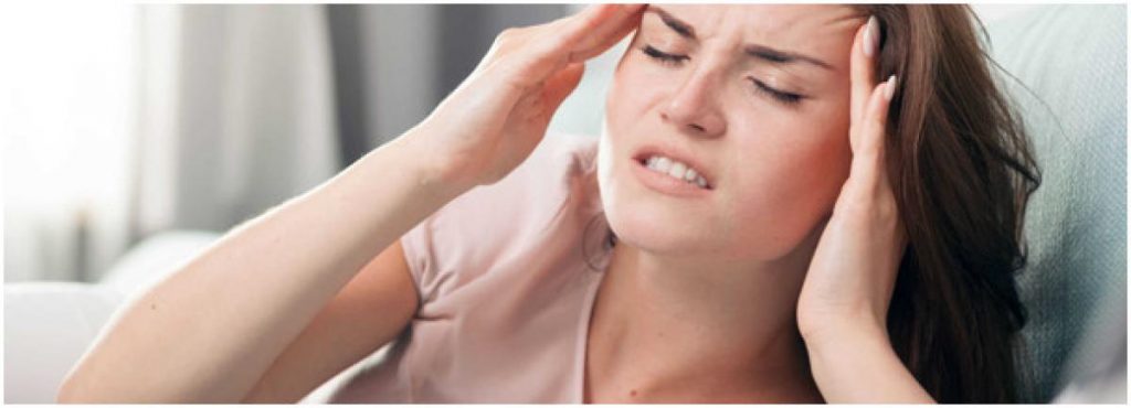 In daily practice, bruxers often seem to have neck pain. During assessment (muscle and joint palpation, physiological and accessory movements), the neck tests may be painful and mobility may be changed. Neck impairments are present in people with TMD and may be one of the risk factors of bruxism (Lobbezoo et al. 2012), but is there a clear relation between cervical dysfunctions/impairments and bruxism? In a recent study, it was noted that the severity of bruxism is independently associated with cervical impairments. Pain associated with cervical movement tests without clear cervical impairments was correlated with bruxism. Therefore, physical therapists need to be aware that signs of cervical movement impairment are not likely to be the cause of bruxism. The coordination is probably related to an increased trigeminal sensitivity which causes false-positive neuromusculoskeletal tests (von Piekartz et al. 2019).
Bruxism is predominantly caused by increased jaw-muscle activity; therefore, muscle orientated treatments like stretching, trigger point treatment, dry needling will have the best outcome
There is evidence that treating the source (masticatory) muscle influences function and reduces pain of the orofacial region (Vázquez Delgado et al. 2010), but there are no clear outcome studies published on muscular treatment modalities in bruxism as defined in the INfORM consensus group in 2013-2018. (Lobbezoo et al. 2013-2018). Because myofascial TMD may be a risk factor for bruxism, increased nociception of the masticatory system may influence the intensity or frequency of bruxism but does not eliminate bruxism (A3 German Guideline Bruxism).
Reflecting on this information, bruxism is a complicated phenomenon that facilitates an interesting debate with many unanswered questions. Bruxism seems to be the domain of the dentist because it has (also) to do with teeth. All credits on research, debates, and consensus go to the dentist which is clearly fair but is it not also the domain of psychology, somnology, and physical therapy? There is a challenge for physical therapy. If physical therapy organizations and researchers execute studies especially on awake bruxism and also integrate and update their knowledge in education for specialized physical therapy, for example, the kind of education CRAFTA provides, can we join the exciting wave dentistry is on? The future will give us the answer.
Do you want to perform the first step? Read the following articles/guidelines for an excellent overview.
- Lobbezoo, F., Ahlberg, J., Raphael, K. G., Wetselaar, P., Glaros, A. G., Kato, T., … & Koyano, K.(2018) International consensus on the assessment of bruxism: Report of a work in progress. Journal of Oral Rehabilitation, 45(11),837-844.
- German Society of Craniomandibular Function and Disorders (DGFDT), German Society of Dental, Oral and Craniomandibular Sciences (DGZMK) S3 Guideline (ect version) Bruxismus Diagnosis and Treatment, J. of Craniomand. Function (2019)11(3):225–i385
Harry von Piekartz
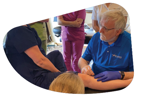
Register for Our Course
Myopain Seminars’ curriculum is developed by international leaders in dry needling and manual trigger point therapy. Our courses are a great way to bring a unique and innovative treatment option to your patients. Completing our course programs elevates your practice and expands your clinician’s toolbox to encompass a wide range of therapies that can effectively eliminate patient pain.
References
- Bader, G., & Lavigne, G. (2000). Sleep bruxism; an overview of an oromandibular sleep movement disorder. Sleep medicine reviews, 4(1), 27-43.
- Cesar, G. M., Tosato, P. J., & Biasotto-Gonzalez, D. A. (2006). Correlation between occlusion and cervical posture in patients with bruxism. Compendium of continuing education in dentistry (Jamesburg, NJ: 1995), 27(8), 463-6.
- Ciancaglini, R., & Radaelli, G. (2001). The relationship between headache and symptoms of temporomandibular disorder in the general population. Journal of dentistry, 29(2), 93-98.
- German Society of Craniomandibular Function and Disorders (DGFDT), German Society of Dental, Oral and Craniomandibular Sciences (DGZMK) S3 Guideline (ect version) Bruxismus Diagnosis and Treatment, J. of Craniomand. Function (2019)11(3):225–i385
- Kritsineli, M., & Shim, Y. S. (1992). Malocclusion, body posture, and temporomandibular disorder in children with primary and mixed dentition. The Journal of clinical pediatric dentistry, 16(2), 86-93.
- Lavigne, G. J., Huynh, N., Kato, T., Okura, K., Adachi, K., Yao, D., & Sessle, B. (2007). Genesis of sleep bruxism: motor and autonomic-cardiac interactions. Archives of oral biology, 52(4), 381-384.
- Lobbezoo, F., & Naeije, M. (2001). Bruxism is mainly regulated centrally, not peripherally. Journal of oral rehabilitation, 28(12), 1085-1091.
- Lobbezoo F, Aarab G, Zaag J van der. Definitions, epidemiology, and etiology of sleep bruxism(2009) In: Lavigne GJ, Cistulli P, Smith M, eds. Sleep medicine for dentists: a practical overview. Chicago: Quintessence Publishing Co, Inc:95–100.
- Lobbezoo, F., van Selms, M., & Naeije, M. (2012)Bruxism and associated risk indicators in Dutch adolescents: a cross-sectional study: O18. Journal of Oral Rehabilitation,39(1).
- Lobbezoo, F., Ahlberg, J., Raphael, K. G., Wetselaar, P., Glaros, A. G., Kato, T., … & Koyano, K.(2018) International consensus on the assessment of bruxism: Report of a work in progress. Journal of oral rehabilitation, 45(11),837-844.
- Manfredini, D., Castroflorio, T., Perinetti, G., & Guarda‐Nardini, L. (2012). Dental occlusion, body posture and temporomandibular disorders: where we are now and where we are heading for. Journal of oral rehabilitation, 39(6), 463-471.
- Manfredini, D., Winocur, E., Guarda-Nardini, L., Paesani, D., & Lobbezoo, F. (2013). Epidemiology of bruxism in adults: a systematic review of the literature. J Orofac Pain, 27(2), 99-110.
- Motta, L. J., Martins, M. D., Fernandes, K. P. S., Mesquita‐Ferrari, R. A., Biasotto‐Gonzalez, D. A., & Bussadori, S. K. (2011). Craniocervical posture and bruxism in children. Physiotherapy Research International, 16(1), 57-61.
- Molina, O. F., dos Santos Jr, J., Nelson, S. J., & Grossman, E. (1997). Prevalence of modalities of headaches and bruxism among patients with craniomandibular disorder. CRANIO®, 15(4), 314-325.
- Muzalev K, Lobbezoo F, Janal MN, Raphael KG.(2017) Inter-episode sleep bruxism intervals and myofascial face pain. Sleep.;40
- Van Selms, M., Lobbezoo, F., Wicks, D. J., Hamburger, H. L., & Naeije, M. (2004). Craniomandibular pain, oral parafunctions, and psychological stress in a longitudinal case study. Journal of oral rehabilitation, 31(8), 738-745.
- von Piekartz, H., Rösner, C., Batz, A., Hall, T., & Ballenberger.(2019)Bruxism, temporomandibular dysfunction and cervical impairments in females–Results from an observational study. Musculoskeletal Science and Practice,45, 102073.
- Paesani DA. Bruxism theory and practice. Chicago: Quintessence Publishing Co, Inc; 2010
- Raphael KG, Janal MN, Sirois DA, et al. (2013)Masticatory muscle sleep background electromyographic activity is elevated in myofascial temporomandibular disorder patients. J Oral Rehabil.;40:883-891.
- Vázquez Delgado, E., Cascos-Romero, J., & Gay Escoda, C. (2010). Myofascial pain associated to trigger points: a literature review. Part 2: differential diagnosis and treatment. Medicina Oral, Patología Oral y Cirugia Bucal, 2010, vol. 15, num. 4, p. 639-643.
